Ningbo Twinkle International Co., LimitedAbout UsRm No.312, Nanyang Mansion ,Yinzhou District,Ningbo, ChinaNingbo Twinkle International Co., LimitedAbout UsRm No.312, Nanyang Mansion ,Yinzhou District,Ningbo, ChinaNingbo Twinkle International Co., LimitedNingbo Twinkle International Co., LimitedNingbo Twinkle International Co., LimitedNingbo Twinkle International Co., LimitedNingbo Twinkle International Co., LimitedNingbo Twinkle International Co., LimitedNingbo Twinkle International Co., LimitedNingbo Twinkle International Co., LimitedNingbo Twinkle International Co., LimitedNingbo Twinkle International Co., LimitedNingbo Twinkle International Co., LimitedNingbo Twinkle International Co., LimitedNingbo Twinkle International Co., LimitedNingbo Twinkle International Co., LimitedNingbo Twinkle International Co., LimitedNingbo Twinkle International

Anatomical Human Eye Model Enlargement Eyeball Orbital Eye Skeleton Optic Nerve Muscles

Anatomical Human Eye Model Enlargement Eyeball Orbital Eye Skeleton Optic Nerve Muscles

Model NO.
LD05015C
Quantity:
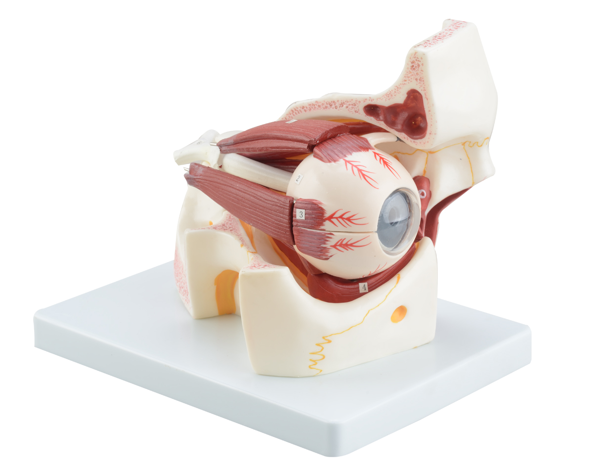
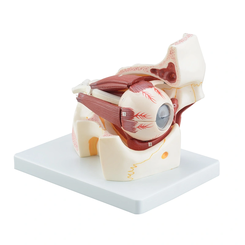
This human eye model shows eye and orbital surrounding structure. The position is under frontal bone, zygomatic above. the 6 muscles can freely change the sight direction, Dissectible into 10 parts, 3 times enlarged. From anterior superficial view, it can help people learn eye muscles, eye nerve, cornea and pupil. From back view, it can help people to learn internal structure of eye including retinal central artery.
This high-quality, life-sized model is perfect for anyone studying or teaching anatomy, optometry, ophthalmology, or neuroscience. It features an enlarged eyeball with realistic details including the cornea, iris, and pupil, and a transparent lens to showcase the inner workings of the eye. The skeletal orbit includes all of the bones of the eye socket, and the optic nerve is fully represented with realistic branching detail. The human eyeball model also includes the six muscles that control eye movements, as well as a stand for convenient display and study. Use this anatomical eye model to demonstrate eye anatomy and function, as well as to diagnose and treat eye disorders.

Send Message

*
* clear input
Preview on Mobile
X
message Message
go top Top
qrcode

Send Message

*
* clear input
Choose language